Effects of Iodine Solution on Histopathologic Features of Lead Acetate-Induced Rat Hepar
Article Sidebar
Main Article Content
Abstract
Background: Lead is a heavy metal that can be found in the environment. Lead can be naturally occurring or produced from human activities. Lead entering the body can cause oxidative stress which can then cause cell damage. Iodine in the body can act as an antioxidant which then prevents oxidative stress.
Methods: This study is an experimental study on 18 Rattus novergicus rats conducted for 19 days. Rats were divided into three groups: negative, positive control group, and treatment group. On the 20th day the rats were killed then the hepatic organs were taken and preparations were made. The preparations were assessed using Knodell’s Score. Data were analyzed using Kruskal-Wallis and then followed by Mann-Whitney Test.
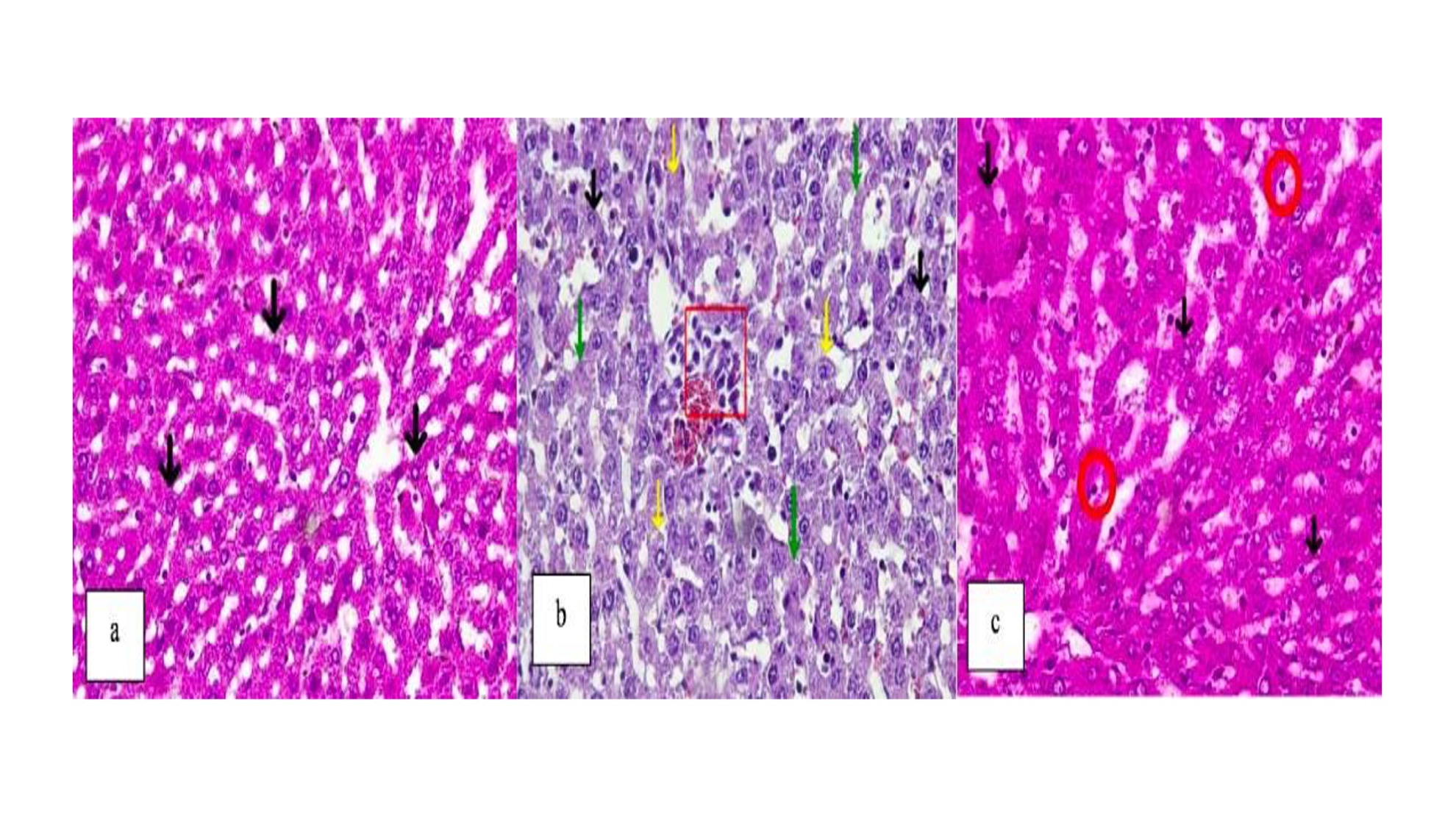
Results: The negative control group showed that the dominant is in the form of normal hepatocytes whilst the positive control group hepatic cells experienced cell damage in the form of inflammation, degeneration, and necrosis. In the treatment group, damage to hepatic cells was lower than the positive control group.
Conclusion: There is an effect of iodine solution administration in preventing damage to rat hepatic cells induced by lead acetate.
Article Details

This work is licensed under a Creative Commons Attribution 4.0 International License.
How to Cite
References
Ismiyati, Marlita D, Saidah D. Pencemaran Udara Akibat Emisi Gas Buang Kendaraan Bermotor. J Manaj Transp Logistik . J Manaj Transp Logistik. 2014;01(03):241–8.
Ardillah Y. Risk Factors for Blood Lead Levels. J Ilmu Kesehat Masy. 2016;7(November):150–5.
Kuswandi. Heavy Metals and Health. 1st ed. Yogyakarta: Grafika Indah; 2017.
Rahman F, Oktomalioputri B, Irramah MI. Effect of Duwet Leaf Extract (Syzigium cumini) on the Histology of Rat Kidney (Rattus novergicus) Intoxicated with Lead Acetate. J Kesehat Andalas. 2020;9(1S):171–7.
Kasanah M, Setiani O, Joko T. Correlation between air lead (Pb) levels and blood lead (Pb) levels among painters in the car body industry in Semarang. J Kesehat Masy. 2016;4(3):825–32.
Fibrianti LD, Azizah R. Characteristics, Blood Lead (Pb) Levels, and Hypertension of Used Battery Home Industry Workers in Talun Village, Sukodadi District, Lamongan Regency. J Kesehat Lingkung. 2015;8(1):92.
Amin I, Hussain I, Rehman MU, Mir BA, Ganaie SA, Ahmad SB, et al. Zingerone prevents lead-induced toxicity in liver and kidney tissues by regulating the oxidative damage in Wistar rats. J Food Biochem. 2021;45(3):1–14.
Suprijono A, Banun S. Effect of Per Oral Lead (Pb) on Histopathological Figures of Hepars a Laboratory Experimental Study In Male Wistar Rats (Rattus Norvegicus). Maj Ilm Sultan Agung. 2017;50(126).
Omotoso BR, Abiodun AA, Ijomone OM, Adewole SO. Lead-Induced Damage on Hepatocytes and Hepatic Reticular Fibres in Rats; Protective Role of Aqueous Extract of Moringa oleifera Leaves (Lam). J Biosci Med. 2015;03(05):27–35.
Mannan R, Misra V, Misra SP, Singh PA, Dwivedi M. A comparative evaluation of scoring systems for assessing necro-inflammatory activity and fibrosis in liver biopsies of patients with chronic viral hepatitis. J Clin Diagnostic Res. 2014;8(8).
Winkler R. Iodine—A Potential Antioxidant and the Role of Iodine/Iodide in Health and Disease. Nat Sci. 2015;07(12):548–57.
Aceves C, Mendieta I, Anguiano B, Delgado-González E. Molecular iodine has extrathyroidal effects as an antioxidant, differentiator, and immunomodulator. Int J Mol Sci. 2021;22(3):1–15.
Vidal ZEO, Rufino SC, Tlaxcalteco EH, Trejo CH, Campos RM, Meza MN, et al. Oxidative stress increased in pregnant women with iodine deficiency. Biol Trace Elem Res. 2014;157(3):211–7.
Choudhry H, Nasrullah M. Iodine consumption and cognitive performance: Confirmation of adequate consumption. Food Sci Nutr. 2018;6(6):1341–51.
Hegazy AMS, Fouad UA. Evaluation of Lead Hepatotoxicity; Histological, Histochemical and Ultrastructural Study. Forensic Med Anat Res. 2014;02(03):70–9.
Thomas VG. Health Risks from Lead-Based Ammunition in the Environment. Ambio. 2013;42(6):737–45.
Fan Y, Zhao X, Wang C, Yu J, Xie J, Li C, et al. Lead-induced oxidative damage in rats / mice : A meta-analysis. J Trace Elem Med Biol. 2019;(December).
Sharma S, Raghuvanshi S, Jaswal A, Shrivastava S, Shukla S. Lead acetate-induced hepatoxicity in wistar rats: Possible protective role of combination therapy. J Environ Pathol Toxicol Oncol. 2015;34(1):23–34.
Wani AL, Ara A, Usmani JA. Lead toxicity: A review. Interdiscip Toxicol. 2015;8(2):55–64.
Fang JY, Wang PW, Huang CH, Hung YY, Pan TL. Evaluation of the hepatotoxic risk caused by lead acetate via skin exposure using a proteomic approach. Proteomics. 2014;14(21–22):2588–99.
Aceves C, Anguiano B, Delgado G. The extrathyronine actions of iodine as antioxidant, apoptotic, and differentiation factor in various tissues. Thyroid. 2013;23(8):938–46.